
In this Article
If you are pregnant, you must be doing everything possible to make sure that your baby gets all the nutrition he needs for his proper growth and development. The thought of having a baby in a couple of months can be overwhelming (and we know you must be a little anxious too). You must be curious to know how your baby is developing and you can – with the help of ultrasound scans. Your doctor will suggest ultrasound scans to check the progress of your baby. An ultrasound is a medical test that produces live images of the foetus using high-frequency sound waves. If you are eager to know how soon you can see your baby on an ultrasound, then read on.
How Early Can You See a Baby on the Ultrasound Scan?
An ultrasound is done every few weeks to assess the growth and well-being of the foetus. The first ultrasound is usually done in the 6th week of pregnancy to detect the gestational sac and to confirm the baby’s position and check the heartbeat of the baby. The gestational sac is seen quite early on before the foetus becomes visible. But of course, you will want to know when you will be able to see the foetus.
By the 6-7th week of conception, fragments of the foetus will be evident on the scan. Your doctor will be able to detect the heartbeat of the foetus by this time. Usually, the heartbeat of the foetus is detected in the 6th week of pregnancy.
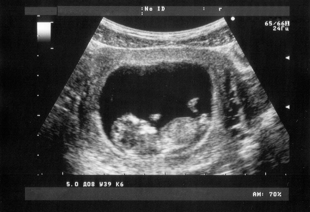
In a week or so, you will be able to see a bean-shaped foetus on the ultrasound scan, and your doctor will probably point out the head end of the baby. However, the small features of the baby can only be seen around 15 weeks. Usually, the ultrasound images are very clear and you will be able to see most of your baby’s features by the 27-28th week of your pregnancy.
What Features of the Baby Can Be Seen on the Ultrasound?
You will be able to notice the following features of your baby on the ultrasound scan –
1. Location
By conducting an ultrasound scan in the sixth week of pregnancy, the doctor is able to tell about the location of your baby. It is during this scan, she will be able to determine whether the baby is in an ideal location or not. She will also be able to rule out the possibility of an ectopic pregnancy (when the embryo attaches to the outside of the uterus) during this scan. But you must note that in some cases that the doctor may not be able to detect the location of the baby in the 6th week and it is completely alright. Every pregnancy is different so you need not worry if the location of your baby is not detected in the 6th week.
2. Heartbeat
As mentioned above, the baby’s heartbeat can be seen on the ultrasound scan by the 6th to 7th week of conception. This confirms that the mother and the baby are in good health and the baby is developing fine. If the heartbeat is not heard or seen, your doctor will suggest another scan in the following week. The heartbeat of the foetus should be around 120-140 beats per minute.
3. Head Ends
The shape of the embryo begins to form around the 8th and 9th week of pregnancy. It is a bean-like shape and the first traces of the complete foetus. The doctor, at this stage, can point out to you the inclination of the baby and its head end.
4. Yolk sac and Chorionic Sac
The chorionic sac refers to the fluid sac in which the baby is carried for 9 months. The yolk sac, on the other hand, is the sac that carries all the nutrients required for the growth of the embryo. These are the earliest to be caught on the ultrasound scan.
Different Types of Ultrasound Scans Performed to See the Baby
The various types of ultrasound scans that help check the progress of the baby are stated below –
1. Standard Ultrasound
This is the regular ultrasound which helps create 2D images of the developing foetus in the womb. In this ultrasound scan, high-frequency sound waves are emitted from a machine to map your body.
2. Doppler Ultrasound
This ultrasound checks the characteristics of the blood flow in the baby’s body. This is vital in case the mother’s blood pressure has been deviant from the health measure. This scan uses high-frequency sound waves to target at the pregnant woman’s blood vessels.
3. Transvaginal Ultrasound
A transvaginal scan helps produce clear images and can be performed in the initial weeks of pregnancy. To perform this scan, the technician inserts a probe about two to three inches into the vaginal passage. This probe is covered with a latex sheath which has gel on it to allow easy entry. A transvaginal ultrasound scan helps obtain clear images with the help of the transducer and helps the doctor detect any abnormalities in the foetus.
4. Foetal Echocardiography
The foetal echocardiography (echo) uses sound waves to check the heart of the foetus. This test helps the doctor detect defects in the heart of the baby before his birth. If any abnormalities are found in the heart, the doctor will be able to provide the treatment soon.
Should You Worry If the Foetus Is Not Visible on the Ultrasound Scan?
The heartbeat and chorionic sac are the two vital indicators of a healthy pregnancy. But another factor which is an indicator of a healthy pregnancy is the size of the embryo. If the size of the embryo is small, it could mean that you have ovulated late. In this case, the due date of the child will be different from a normal 9 months. The baby may also not be visible at 6 weeks and thus you may have to visit the doctor multiple times.
If there is no sign of heartbeat after subsequent check-ups after 8 weeks, then it could be a sign of an ectopic pregnancy or even a miscarriage.
If you are pregnant for the first time, we suggest that you go for each and every ultrasound suggested by your doctor. Ultrasound scans are important to assess the health and the development of the foetus. You have no reason to be afraid of ultrasound scans, they are harmless and don’t result in pain. So go for all your scans and see how your baby is developing!


